What Is a Cavity Between Teeth?
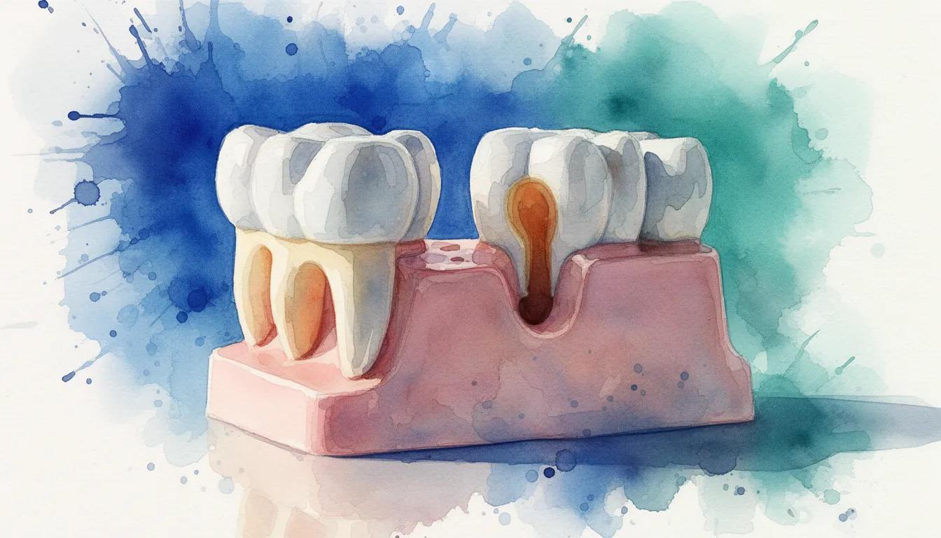
An interproximal cavity is tooth decay that forms on the surface where two teeth touch each other. These cavities develop in the narrow gap between teeth, a space that is difficult to clean and easy to overlook.
Dental caries, the clinical term for cavities, is one of the most common chronic diseases worldwide. A 2022 study in the Journal of Dental Research found that untreated dental caries in permanent teeth affected roughly 2 billion people globally as of 2019, making it a persistent public health challenge. [3] The interproximal surface is one of the most frequent sites where this decay begins.
Interproximal cavities are especially common in the premolars and molars, the back teeth that sit close together. The tight contact point between these teeth traps food particles and plaque, a sticky film of bacteria. Over time, the bacteria produce acids that dissolve tooth enamel, the hard outer layer of the tooth. Because this process happens in a hidden area, the cavity can grow substantially before you or your dentist notice it during a routine visual check.
Children and adults are both affected. Systematic reviews have shown high prevalence of dental caries in both primary (baby) and permanent teeth across all age groups worldwide. [7] Understanding where and why these hidden cavities form is the first step toward catching them early.
Causes and Risk Factors for Interproximal Cavities
Interproximal cavities are caused by acid-producing bacteria in plaque that collect between teeth where brushing alone cannot reach.
How Decay Starts Between Teeth
Plaque builds up on every tooth surface, but it is hardest to remove from the contact area between teeth. When you eat sugary or starchy foods, bacteria in plaque convert those sugars into acids. These acids attack the enamel, pulling out minerals in a process called demineralization. If the minerals are not replaced through saliva and fluoride, a cavity forms.
The contact point between two teeth is especially vulnerable. Toothbrush bristles are too wide to fit into this narrow gap. Without flossing or another interdental cleaning method, plaque sits undisturbed in this space for hours or days, giving bacteria plenty of time to cause damage.
Diet and Oral Hygiene Habits
Frequent snacking on sugary or acidic foods feeds the bacteria that cause decay. Sipping on sweetened drinks throughout the day keeps acid levels high in the mouth. Each acid exposure can last about 20 to 30 minutes after eating.
Skipping daily flossing is one of the biggest risk factors for interproximal cavities specifically, because the contact area between teeth is the one surface a toothbrush cannot clean. According to the American Dental Association, cleaning between teeth once a day is essential for removing plaque from surfaces a toothbrush misses. [12] However, preventing cavities overall requires a combination of approaches: brushing with fluoride toothpaste, flossing daily, limiting sugar intake, and visiting the dentist regularly. The CDC emphasizes that no single method works in isolation. [13]
- Frequent sugar consumption throughout the day
- Acidic beverages such as soda, citrus juice, or sports drinks
- Inconsistent or absent flossing routine
- Dry mouth (xerostomia), which reduces saliva's natural cleansing and remineralizing ability
Other Contributing Factors
Crowded or overlapping teeth create extra-tight contact points that trap more plaque. Orthodontic appliances like braces can also make interproximal cleaning harder. Certain medical conditions that reduce saliva flow, such as Sjögren's syndrome or medication side effects, raise cavity risk across all tooth surfaces.
Teeth with structural defects, such as molar-incisor hypomineralization (MIH), a condition where enamel forms with less mineral content, are more vulnerable to caries. Research has identified MIH as a significant risk factor for decay, particularly in children. [5] [2] These weakened enamel areas break down faster when exposed to bacterial acids.
Symptoms and Diagnosis
Early interproximal cavities usually cause no symptoms at all, which is why routine X-rays are critical for detection.
What Patients Experience
In the earliest stage, you may notice nothing. The enamel breakdown is too small to see or feel. As the cavity grows deeper, common symptoms include sensitivity to hot, cold, or sweet foods and drinks. You may feel a sharp twinge when biting down on food in that area.
When the decay reaches the dentin, the softer layer beneath enamel, sensitivity typically increases. Food may get stuck more easily between the affected teeth. If the cavity advances further and reaches the pulp, the nerve-containing tissue inside the tooth, you may experience a persistent toothache, throbbing pain, or swelling. At this stage, the damage is significant and often requires more than a simple filling.
- No symptoms in early stages
- Mild sensitivity to temperature or sweetness as enamel thins
- Food catching or floss shredding between specific teeth
- Visible dark spot or hole (usually only when the cavity is already large)
- Persistent or spontaneous pain if decay reaches the pulp
How Dentists Detect Interproximal Cavities
Bitewing X-rays are the standard diagnostic tool. These are small X-ray images taken while you bite down on a tab, capturing the contact areas of your upper and lower back teeth on one film. A cavity shows up as a dark shadow in the enamel or dentin between teeth.
A 2021 systematic review and meta-analysis published in Caries Research evaluated various detection methods for early caries. The review found that while visual-tactile examination is commonly used, radiographic methods, particularly bitewing radiography, remain essential for identifying interproximal lesions that are not visible to the naked eye. [6] Some dental offices also use digital sensors, laser fluorescence devices, or near-infrared transillumination as supplementary tools, though these are not yet a universal replacement for X-rays.
The American Dental Association recommends that the frequency of bitewing X-rays be based on individual risk. [12] If you have a history of cavities, your dentist may recommend X-rays every six to twelve months. Lower-risk patients may need them less often.
Treatment Options for Cavities Between Teeth
Treatment depends on how deep the cavity has progressed, ranging from fluoride therapy for very early lesions to root canal treatment for advanced decay.
Fluoride and Remineralization for Very Early Decay
When a cavity is caught at the earliest stage, before it breaks through the enamel surface, it may be possible to reverse the damage without drilling. This stage is sometimes called a "white spot lesion" or incipient caries. Professional fluoride varnish or prescription-strength fluoride toothpaste can help remineralize the weakened enamel.
A Cochrane systematic review found that fluoride toothpaste with a concentration of 1,000 parts per million (ppm) or higher significantly reduced dental caries in both children and adolescents compared to placebo. [9] This evidence supports using fluoride toothpaste as a daily preventive measure and, in early decay, as part of a remineralization strategy. Your dentist may also recommend a high-fluoride prescription rinse or gel for high-risk patients.
Dental Fillings
Once the cavity has broken through the enamel into the dentin, a filling is typically needed. The dentist removes the decayed tooth structure and replaces it with a filling material. Composite resin, a tooth-colored material, is the most common choice for interproximal fillings because it bonds directly to the tooth and blends in visually.
Placing a filling between teeth requires careful technique. The dentist uses a thin metal band called a matrix band to shape the filling material so it recreates the natural contour of the tooth wall. A well-placed filling restores the contact point between the two teeth, which helps prevent food trapping in the future.
Inlays, Onlays, and Crowns
If the cavity is large and has removed a significant amount of tooth structure, a standard filling may not be strong enough. In these cases, an inlay or onlay, which are custom-made restorations fabricated in a dental laboratory, may be a better option. An inlay fits inside the cusps (the raised points) of a tooth. An onlay covers one or more cusps.
When the cavity has destroyed most of the visible tooth, a crown may be necessary. A crown is a cap that covers the entire visible portion of the tooth. It restores the tooth's shape, strength, and function. Crowns can be made from porcelain, metal alloys, or a combination of both.
Root Canal Treatment
When an interproximal cavity reaches the pulp, the soft tissue inside the tooth that contains nerves and blood vessels, a filling alone will not solve the problem. The infected pulp must be removed to save the tooth. This procedure is called root canal treatment, or endodontic therapy.
During root canal treatment, an endodontist, a dentist who specializes in treating the inside of the tooth, removes the infected pulp tissue, cleans and shapes the root canals, and fills them with a biocompatible material. A systematic review and meta-analysis on vital pulp therapy for deep caries found that preserving pulp vitality through techniques such as partial or complete caries removal can be effective in certain cases, particularly in immature teeth. [4] However, when the pulp is irreversibly inflamed or infected, root canal treatment becomes the necessary step to eliminate infection and preserve the tooth.
After root canal treatment, the tooth typically needs a crown to protect it from fracture. You can learn more about when and why you might need to see this type of specialist on the endodontics page.
Tooth Extraction as a Last Resort
If the cavity has destroyed too much tooth structure to restore, or if infection has spread beyond what root canal treatment can resolve, extraction may be the only remaining option. After extraction, your dentist or specialist will discuss replacement options such as a dental implant, bridge, or removable partial denture to restore function and prevent neighboring teeth from shifting.
Recovery and Aftercare
Recovery time depends on the treatment you receive, but most patients resume normal eating within a day or two after a filling.
After a Filling, Inlay, or Crown
Sensitivity after a filling is common and usually fades within a few days to two weeks. Avoid very hot or cold foods during this period. If you received a local anesthetic, wait until the numbness wears off before eating to avoid biting your cheek or tongue.
For inlays, onlays, or crowns that require laboratory fabrication, you may wear a temporary restoration for one to three weeks. Be gentle when flossing around the temporary. Once the permanent restoration is placed, brush and floss normally. Your dentist will check your bite and adjust if needed.
- Mild sensitivity for a few days to two weeks is typical after a filling
- Temporary restorations require gentle cleaning; avoid sticky foods
- Return for your follow-up appointment so the dentist can verify the restoration's fit
After Root Canal Treatment
Mild soreness near the treated tooth is normal for two to five days after a root canal. Over-the-counter pain relievers such as ibuprofen typically manage this discomfort well. The American Association of Endodontists notes that most patients return to their normal activities the next day. [11]
Avoid chewing on the treated tooth until the permanent crown is placed. A tooth that has had root canal treatment without a crown is more prone to fracture. Keep up with your regular brushing and flossing routine, and attend all scheduled follow-up visits.
Preventing Future Interproximal Cavities
The best long-term strategy combines daily flossing, fluoride toothpaste with at least 1,000 ppm fluoride, regular dental checkups with bitewing X-rays, and a diet low in frequent sugar exposure. [9] [12] [13] Limiting snacking between meals and choosing water over sugary drinks also reduces acid attacks on your teeth. No single preventive measure works well on its own. The strongest protection comes from using all of these approaches together.
If you are at high risk for cavities, your dentist may recommend prescription-strength fluoride toothpaste (5,000 ppm), in-office fluoride varnish treatments, or antimicrobial rinses. Sealants, while typically used on biting surfaces, are not practical for interproximal areas, so flossing remains your primary defense between teeth.
Cost Factors for Treating Interproximal Cavities
The cost of treating a cavity between teeth varies widely based on the treatment type, location, provider, and case complexity.
A composite filling for an interproximal cavity typically ranges from $150 to $400 per tooth. Inlays and onlays, because they require laboratory fabrication, generally cost between $500 and $1,200. A full crown may range from $800 to $1,800 or more depending on the material chosen and geographic region. Costs vary by location, provider, and case complexity.
Root canal treatment costs depend on which tooth is involved. Front teeth, with a single root canal, are typically less expensive than molars, which have three or four canals. Root canal treatment on a molar may range from $900 to $1,500 or more, not including the cost of the crown that usually follows. Again, costs vary by location, provider, and case complexity.
Most dental insurance plans cover a portion of fillings, crowns, and root canal treatments, though coverage levels differ. Fillings are often covered at 70% to 80% under a basic restorative benefit. Crowns and root canals may fall under major restorative, typically covered at 50% to 60%. Check with your insurance provider for specific plan details. Many dental offices also offer payment plans or accept third-party financing to help spread costs over time.
When to See a Specialist
Most interproximal cavities are treated by a general dentist, but advanced decay that reaches the pulp often requires an endodontist.
A general dentist handles the majority of fillings, inlays, onlays, and crowns. When the cavity is deep enough to involve the nerve of the tooth, or when initial root canal treatment does not fully resolve an infection, your dentist may refer you to an endodontist. Endodontists complete two or more additional years of training beyond dental school, focused specifically on diagnosing and treating problems inside the tooth. [11]
You may also need a referral if your tooth has unusual root anatomy, a cracked root, or persistent pain after a previous root canal. Endodontists use specialized tools such as dental operating microscopes and cone-beam computed tomography (CBCT, a type of 3D X-ray) to see structures that standard X-rays may miss.
If your general dentist identifies a deep interproximal cavity and suspects pulp involvement, asking about a referral to an endodontist is a reasonable next step. Early referral can improve the chances of saving the tooth. You can find more information about this specialty on the endodontics page.
Find a Specialist Near You
If you have been told you have a deep cavity between your teeth, or if you are experiencing persistent tooth pain, connecting with a qualified endodontist can help you understand your options. Use the My Specialty Dentist directory to search for an endodontist in your area, read about their training and experience, and take the next step toward getting the care your tooth needs.
Search Endodontists in Your Area