Overview: Digital vs Film Dental X-Rays
Digital X-rays use an electronic sensor to capture an image on a computer, while film X-rays use a small sheet of coated plastic that must be developed in chemicals. Both technologies produce diagnostic dental radiographs [1].
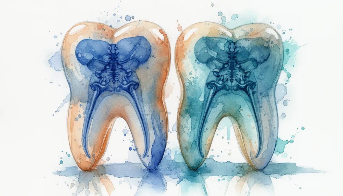
Dental X-rays, also called radiographs, let your dentist see structures that are hidden from a visual exam. These include the inside of teeth, the roots, the bone that supports the teeth, and developing teeth in children [2]. The X-ray itself is the same form of energy whether it strikes a film or a digital sensor. The difference is in how the image is recorded and viewed.
Film radiography was the standard in dentistry for most of the twentieth century. Digital systems began entering practices in the late 1980s and have become the more common choice in many offices today [1]. Some practices still use film, and some use both depending on the exam.
For most patients, the choice between digital and film is made by the office, not the patient. Understanding the differences helps you ask better questions about radiation, image quality, and how your records are stored.
How Each Technology Works
Both systems use the same X-ray beam, but they record the image in different ways. Film uses a chemical reaction on a silver-coated sheet, while digital uses an electronic sensor that converts X-ray energy into a digital signal [1].
How Film X-Rays Work
A small film packet is placed inside your mouth. The X-ray machine sends a short burst of radiation through your tooth and jaw. The radiation that passes through hits the film and exposes the silver crystals on its surface.
The exposed film is then developed in a series of chemical baths in a darkroom, similar to old photographic film. After development, the image appears as a physical picture that the dentist holds up to a light box to read. Film images cannot be changed after they are processed. If the exposure was off, the X-ray must be retaken.
How Digital X-Rays Work
A digital sensor takes the place of the film. The two main types are direct sensors, which connect to a computer by a cable, and indirect sensors called photostimulable phosphor plates, which are scanned after the exposure [1].
When the X-ray beam strikes the sensor, the energy is converted into electrical signals that the computer turns into an image. The image appears on a monitor within seconds. Software lets the dentist zoom in, change brightness and contrast, and measure distances on the image [1].
What Dental X-Rays Are Used For
Dental X-rays help diagnose cavities, gum disease, infections, bone loss, and problems that are not visible during a regular exam. Both digital and film are used for the same clinical purposes [2].
Routine Checkup Imaging
Bitewing X-rays show the crowns of the upper and lower teeth in one image. They help dentists find cavities between teeth and check the height of the bone around them [2]. Periapical X-rays show the entire tooth from crown to root tip and the surrounding bone. They are used to evaluate root infections, abscesses, and changes in bone.
How often these images are taken depends on your age, risk for cavities, gum health, and history of dental problems. The American Dental Association recommends that the frequency of X-rays be based on individual need, not a fixed schedule [2].
Specialty and Surgical Applications
Panoramic X-rays capture the entire mouth in a single image, including all teeth, the upper and lower jaws, and parts of the sinuses and jaw joints. They are commonly used before wisdom tooth removal, for orthodontic planning, and to evaluate jaw fractures or cysts.
Cone beam computed tomography, or CBCT, is a three-dimensional imaging technique used for implant planning, evaluation of impacted teeth, and assessment of complex root canal anatomy. CBCT is a separate technology from standard digital X-rays and uses a higher radiation dose than two-dimensional images [1]. Learn more on the oral-radiology page.
Evidence and Diagnostic Performance
Research generally shows that digital and film X-rays produce similar diagnostic results when both are exposed and processed correctly. The American Academy of Oral and Maxillofacial Radiology and the American Dental Association both recognize digital radiography as an accepted standard in dental practice [1] [2].
Regulatory Status
Dental X-ray sensors and digital imaging systems sold in the United States are cleared by the Food and Drug Administration as medical devices. FDA clearance means the device has been shown to be substantially equivalent to other devices already on the market. It is not the same as FDA approval, which applies to a smaller category of higher-risk devices.
X-ray equipment is also regulated by state agencies, which require inspections and certified operators.
Professional Society Positions
The American Dental Association and the American Academy of Oral and Maxillofacial Radiology support the use of dental X-rays for diagnosis and treatment planning, and they recommend using the lowest reasonable dose to achieve a useful image. This principle is called ALARA, which stands for As Low As Reasonably Achievable [1] [2].
Both organizations emphasize that the choice between digital and film should be based on the practice's setup, the clinician's training, and the diagnostic task, not on marketing claims. In many cases, digital is preferred because of lower dose and easier workflow, but well-exposed film can produce diagnostic images of comparable quality [1].
Benefits and Limitations
Both digital and film have real strengths and weaknesses. The right choice depends on the practice setting, the type of exam, and the specific question the dentist is trying to answer.
Advantages of Digital X-Rays
Digital sensors are more sensitive to X-rays than traditional film. This generally allows the dentist to use a shorter exposure, which reduces the radiation dose to the patient. Reductions of roughly half or more compared to film are commonly reported, although the exact amount depends on the sensor type and technique [1].
Other practical advantages include:
- Images appear on the screen within seconds, so the dentist can review them during your visit.
- Images can be zoomed, brightened, or contrast-adjusted on the monitor, which may reduce the need for retakes.
- Files can be stored on a computer or in the cloud and shared with specialists by secure email or referral portal.
- No darkroom, chemicals, or chemical disposal is required.
- Easier integration with electronic health records and treatment planning software.
Advantages of Film X-Rays
Film has a long track record in dentistry and does not depend on a computer system or sensor that can fail. Initial equipment costs for a film setup are typically lower than for a full digital system, although operating costs over time can be higher because of film, chemicals, and processing equipment.
Some clinicians feel that high-resolution film offers very fine detail in specific situations. In day-to-day diagnostic use, the difference is rarely meaningful when both are used correctly [1].
Limitations to Be Aware Of
Digital sensors are bulkier and stiffer than film packets. Some patients, especially those with small mouths or strong gag reflexes, find them less comfortable. Phosphor plate systems are thinner and more flexible but require an extra scanning step.
Film requires careful handling, controlled developing chemicals, and physical storage space. Lost or damaged film cannot be recovered. Improper processing can produce images that are too dark, too light, or unreadable, which may require retakes and additional radiation exposure.
Cost and Availability
Patients typically pay a similar amount for a dental X-ray exam whether the office uses digital or film, because billing is based on the type and number of images, not the technology used. Costs vary by location, provider, and case complexity.
What Patients Typically Pay
Without insurance, a single bitewing or periapical X-ray often falls in the range of $25 to $50 in many areas. A full mouth series of about 18 images often falls between $100 and $250. A panoramic X-ray typically ranges from $60 to $150. A CBCT scan, which is a separate three-dimensional study, often ranges from $150 to $500 depending on the field of view. These ranges are general and vary by location, provider, and case complexity.
Most dental insurance plans cover routine X-rays at recommended intervals, often once or twice per year for bitewings and less frequently for full mouth series or panoramic images. Coverage for CBCT varies and is sometimes considered a separate medical or specialty service. Check your specific plan benefits before treatment.
Finding the Technology in Your Area
Digital X-ray systems are widely available across the United States. Many general dentists and most dental specialists use digital systems for routine intraoral imaging and panoramic exams [1]. CBCT is more common in oral surgery, endodontics, periodontics, and orthodontic practices.
If digital imaging matters to you, ask the office directly when you schedule. Some practices use a mix, with digital for intraoral exams and a separate panoramic or CBCT system.
Questions to Ask Your Dentist
A short, direct conversation with your dentist or hygienist is the best way to understand what imaging will be used and why. The goal is informed care, not avoiding necessary X-rays.
- Do you use digital or film for intraoral X-rays?
- Why are these specific X-rays needed today?
- How often do you recommend X-rays for someone with my history and risk?
- What dose-reduction practices do you use, such as collimation and lead aprons or thyroid collars where appropriate?
- Can my images be shared electronically with a specialist if I am referred?
- If I had X-rays at another office recently, can those be used instead of new ones?
- If I am pregnant or might be pregnant, how will that change your plan?
Find an Oral Radiology Specialist
Most general dentists handle routine X-rays well. For complex imaging needs such as CBCT interpretation, suspected jaw pathology, or pre-surgical planning, an oral and maxillofacial radiologist or another dental specialist can help. Browse the oral-radiology page to learn more and find a specialist near you.
Search Oral Radiologists in Your Area