What Is Laser Cavity Detection?
Laser cavity detection is a chairside method that uses a low-power laser to find early tooth decay before it forms a visible hole. The device measures fluorescence, a soft glow given off by bacterial byproducts trapped inside damaged enamel and dentin[2].
The most widely known unit is DIAGNOdent, made by KaVo. A handheld pen tip touches the tooth, sends a focused beam of red light into the surface, and reads the light that comes back. Healthy enamel reflects very little fluorescence. Decayed enamel reflects more, and the device translates that difference into a number on a small display[6].
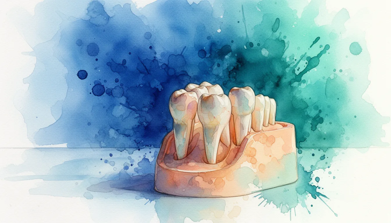
Dentists and endodontists use laser detection alongside the tools they already trust: a sharp eye, a mirror, an explorer, and bitewing radiographs. The laser is a second opinion in a region of the tooth that hides decay well, the chewing surface, where deep grooves can mask trouble for years.
How the Technology Works
The device works by detecting a specific wavelength of light that decayed tooth structure gives off when stimulated by a red laser. The signal comes from porphyrins, organic compounds produced by the bacteria that cause cavities[2].
The DIAGNOdent unit emits laser light at 655 nanometers, in the red part of the visible spectrum. That light enters the tooth and excites the porphyrin molecules sitting in micro-pores left behind by demineralization. Those molecules then emit longer-wavelength fluorescence that travels back up the same fiber tip to a detector[2][5].
Several research groups have measured how light moves through carious versus healthy tooth structure. Carious tissue shows different optical and thermal properties because demineralization changes how enamel scatters and absorbs light[5]. That difference is what makes fluorescence-based devices possible in the first place.
Newer optical methods build on this same principle. Near-infrared transillumination, photothermal imaging, and Raman spectroscopy all try to read changes in the tooth without cutting into it[4][5][10]. Laser fluorescence remains the most familiar of these to general dentists and patients.
Reading the Numerical Scale
The device displays two numbers: a peak value and a moment-to-moment value as the probe moves across the tooth. Clinicians typically interpret the peak reading in ranges. Low numbers suggest healthy or only slightly demineralized tissue. Mid-range numbers suggest enamel decay that may warrant closer monitoring or preventive treatment. Higher numbers suggest decay that has entered dentin and likely needs restoration[6].
Cut-off values are not universal. They depend on the manufacturer's calibration, the tooth surface being scanned, and how the clinician was trained[6]. Because of that, the score is one input, not a verdict.
Clinical Applications
Dentists use laser cavity detection to screen tooth surfaces that are difficult to read with eyes and X-rays alone. It is most valuable in the deep grooves of back teeth and during follow-up of suspicious spots over time.
Occlusal Pits and Fissures
Chewing surfaces of molars and premolars have deep grooves that can hide decay under intact-looking enamel. Fluorescence devices can pick up changes in these grooves that an explorer or visual check would miss[4][6].
This makes the technology especially useful for children, teens, and adults at higher caries risk, where a delayed diagnosis on a back tooth often means a deeper restoration later.
Monitoring Suspicious Spots Over Time
When a dentist sees a stained groove or a faint shadow on an X-ray, the question is often: is this active decay or an old, stable mark? Repeated laser readings at recall visits can show whether a lesion is progressing, holding steady, or remineralizing with fluoride and good home care[4].
Restorative and Endodontic Use
Inside a tooth that is already being treated, clinicians need to know when they have removed enough decayed dentin. A 2024 randomized clinical trial compared methods for detecting residual carious dentin after cavity preparation and found measurable differences between traditional and adjunctive approaches[3].
Endodontists, who treat the soft pulp inside teeth, sometimes work alongside general dentists when deep decay raises the question of whether a tooth can keep its nerve. Reading more about the endodontics page can help you understand when a specialist visit becomes part of the plan.
Evidence and Effectiveness
Research supports laser fluorescence as a useful adjunct, particularly on chewing surfaces, while showing that no single method finds every cavity reliably on its own.
FDA Status
DIAGNOdent and similar laser caries detection devices are marketed in the United States under FDA clearance, the regulatory pathway for devices considered substantially equivalent to existing products. Clearance is not the same as FDA approval, which is reserved for new high-risk devices that must demonstrate safety and effectiveness through more rigorous review. Patients should ask their dentist about the specific device being used and how it is being applied.
What Studies Show
A 2024 systematic review of near-infrared technology for caries detection found that optical methods can identify early lesions, with diagnostic performance varying by tooth surface and lesion depth[4]. A 2022 systematic review and meta-analysis on emerging technologies for dentin caries detection reached similar conclusions: these tools add value, but performance depends on the clinical setting and the comparison standard used[6].
A 2020 in vitro comparison of methods for initial proximal caries detection examined how laser fluorescence and other approaches perform on the between-tooth surfaces that bitewing X-rays usually screen[9]. The findings reinforced that proximal lesions remain harder to read optically than occlusal ones.
Newer optical approaches, including Raman spectroscopy and photothermal imaging, are being studied as the next step beyond fluorescence[5][10]. These remain primarily research tools at this stage and are not standard chairside equipment in general practice.
Professional Society Guidance
Major dental organizations, including the American Dental Association and the American Association of Endodontists, publish patient-facing guidance on diagnostic technology and treatment planning[11][12]. The consistent theme: diagnostic tools should be used in combination, not in isolation, and treatment decisions should rest on a clinician's full assessment.
Benefits and Limitations
Laser cavity detection offers real advantages over visual-only screening, but it also has well-documented blind spots that patients should understand.
Advantages
The scan is painless and quick. There is no needle, no drill, no biting on a film. Each tooth takes only seconds to read once the probe is set up[2].
There is no ionizing radiation. Unlike bitewing X-rays, laser fluorescence uses visible light, so it can be repeated at follow-up visits without radiation dose concerns[2].
Earlier detection on chewing surfaces can support minimally invasive care. When a lesion is found while still in enamel, options like fluoride varnish, sealants, and improved home care may stop or reverse the process before a filling is needed[2][6].
Limitations
False positives are a known issue. Stained grooves, food debris, calculus, plaque, and certain prophy pastes can all raise the reading without true decay present[6]. A high number alone is not a cavity diagnosis.
False negatives also happen. Deeper proximal lesions, decay under existing fillings, and early demineralization in smooth surfaces are not where laser fluorescence shines. X-rays remain essential for these areas[4][9].
Operator technique matters. The probe angle, tip cleanliness, and how the tooth is dried all influence the reading. Calibration at the start of each appointment is part of standard use[6].
The device does not replace clinical judgment. It is a number that a trained clinician interprets in context, alongside the visual exam, X-rays, the patient's caries risk profile, and prior records.
Cost and Availability
Patients usually do not pay a separate fee for a laser scan; offices that own the device typically include it as part of a comprehensive exam. Costs vary by location, provider, and case complexity.
Some practices bill an additional fee for laser-assisted screening, often in the range of a small adjunct charge per visit. Dental insurance plans generally do not list laser caries detection as a covered procedure code, so any extra cost is often out of pocket. Before treatment, ask the office whether the scan is included in the exam fee or billed separately.
Availability has grown as practices have adopted optical caries detection over the past two decades, but the technology is not universal. Pediatric dentists, general dentists with a strong prevention focus, and some endodontists are more likely to have it. Calling ahead is the fastest way to confirm.
Questions to Ask Your Dentist
If laser cavity detection matters to you, a short conversation at your next visit can clarify how the technology fits into your care.
- Do you use a laser fluorescence device such as DIAGNOdent in your exams?
- Is the scan included in the exam fee, or is there a separate charge?
- How do you decide when to treat versus monitor a borderline reading?
- Will you record the readings so we can compare them at future visits?
- How does the laser reading change your treatment plan compared with X-rays alone?
- What home-care steps would you recommend if a reading is elevated but not yet decay?
Find a Specialist
If you have been told a tooth has deep decay near the nerve, or if you want a second opinion on a borderline lesion before drilling, an endodontist can help you weigh the options. Learn more about specialty care on the endodontics page and search the directory to find a specialist near you.
Search Endodontists in Your Area