What Is a Cone Beam CT Scan?
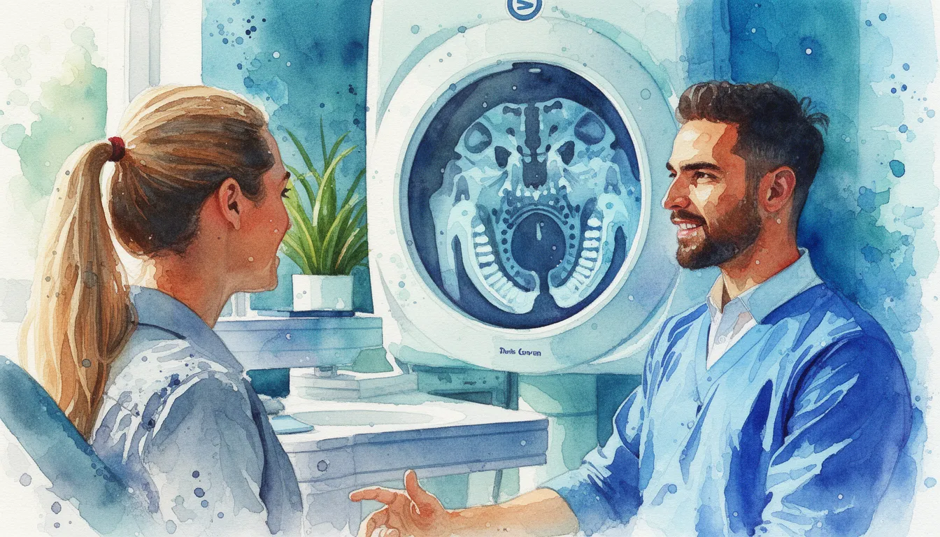
A cone beam computed tomography (CBCT) scan is a specialized type of X-ray that uses a cone-shaped beam to capture a three-dimensional image of your teeth, jawbone, nerve pathways, and soft tissues in a single rotation around your head. Unlike a standard dental X-ray, which produces a flat, two-dimensional image, CBCT creates a detailed 3D model that can be viewed from any angle.
CBCT technology was introduced to dentistry in the late 1990s and has become increasingly common in specialty dental practices. The machine looks similar to a panoramic X-ray unit. You stand or sit while the scanner rotates around your head, capturing hundreds of images that a computer assembles into a three-dimensional volume.
The key advantage of CBCT is precision. It allows your dental provider to see the exact position, size, and shape of anatomical structures that would overlap or be hidden on a traditional X-ray. This level of detail is essential for planning procedures like dental implants, evaluating complex root anatomy, and diagnosing jaw pathology.
When and Why a CBCT Scan Is Ordered
A CBCT scan is not a routine screening tool. It is ordered when your provider needs specific three-dimensional information that a standard X-ray cannot provide. The guiding principle is that the clinical benefit of the scan must outweigh the radiation exposure.
Common Reasons for a CBCT
- Dental implant planning: CBCT shows exact bone width, height, and density at the implant site. It also reveals the location of the inferior alveolar nerve, maxillary sinus, and adjacent tooth roots to prevent complications.
- Impacted teeth: For wisdom teeth or other impacted teeth, CBCT shows the precise relationship between the tooth and nearby nerves, sinuses, and adjacent roots.
- Complex endodontic (root canal) cases: CBCT can reveal additional root canals, root fractures, and the extent of infection that a standard X-ray may miss.
- TMJ disorders: The scan shows the bony components of the temporomandibular joint in detail, helping to identify arthritis, condylar fractures, or structural abnormalities.
- Jaw pathology: Cysts, tumors, and other lesions in the jaw can be precisely measured and their boundaries mapped.
- Orthodontic treatment planning: CBCT can assess impacted canines, root resorption, airway dimensions, and bone support for complex orthodontic cases.
- Trauma evaluation: Jaw fractures, tooth fractures, and displaced teeth can be visualized in 3D.
CBCT vs. Standard Dental X-rays
Standard dental X-rays (periapical and bitewing images) are excellent for detecting cavities, basic bone loss, and routine dental problems. They use very low radiation and remain the workhorse of dental imaging. A panoramic X-ray provides a broader view but is still two-dimensional and can distort sizes and distances.
CBCT fills the gap when two-dimensional images are not enough. It eliminates the overlap of structures that makes flat images hard to interpret in complex situations. However, CBCT is not a replacement for standard X-rays in routine dental care.
What to Expect During a CBCT Scan
A CBCT scan is a quick, painless procedure that takes place in your dentist's or specialist's office. No needles, no contrast dye, and no sedation are involved.
Before the Scan
There is usually no special preparation required. You will be asked to remove any metal objects from the head and neck area, including earrings, necklaces, hearing aids, eyeglasses, and removable dental appliances. Metal creates artifacts on the scan that can interfere with image quality.
If you are pregnant or think you might be, tell your provider before the scan. While CBCT uses less radiation than a medical CT, radiation exposure during pregnancy should be minimized.
During the Scan
You will stand or sit with your chin resting on a small support. A head stabilizer keeps you in the correct position. The CBCT machine has a rotating arm that circles around your head once. The entire scan takes between 10 and 40 seconds, depending on the area being imaged and the machine model.
You must remain completely still during the scan. Any movement can cause blurring in the images. Breathing normally is fine. The machine makes a quiet whirring sound as it rotates. There is no discomfort, no enclosed space, and no injection.
After the Scan
The scan data is processed by the computer within minutes. Your provider can view the 3D images almost immediately. In some offices, the images are interpreted on-site. In others, particularly for complex cases, the images may be sent to an oral and maxillofacial radiologist for a formal report. You can return to normal activities immediately after the scan.
Radiation Safety and Image Interpretation
One of the most common questions patients have about CBCT is radiation exposure. Understanding the actual dose and how it compares to other sources helps put the risk in perspective.
How Much Radiation Does a CBCT Use?
The radiation dose from a dental CBCT scan typically ranges from 20 to 200 microsieverts (uSv), depending on the field of view and machine settings. For comparison, a single standard dental X-ray delivers about 5 uSv. A panoramic X-ray delivers about 10-25 uSv. A full medical CT of the head delivers about 2,000 uSv.
This means a CBCT scan uses roughly 4 to 40 times more radiation than a single dental X-ray, but only about 1/10th to 1/100th the dose of a medical CT scan. The average person receives about 3,000 uSv per year from natural background radiation (cosmic rays, radon, etc.).
The ALARA Principle
All dental imaging follows the ALARA principle: As Low As Reasonably Achievable. This means your provider should only order a CBCT when the diagnostic benefit justifies the radiation dose. Good practice includes using the smallest possible field of view (imaging only the area of interest rather than the entire head) and using the lowest dose settings that still produce diagnostically useful images.
Who Reads Your CBCT Scan?
Any dentist who orders a CBCT has an obligation to review the entire scan volume, not just the area of interest. This includes checking for incidental findings in the sinuses, airway, cervical spine, and other structures that appear in the scan. An oral and maxillofacial radiologist is the specialist best trained to interpret these images and identify findings that a treating dentist might overlook.
Cost Factors for a CBCT Scan
Costs vary by location and provider. Several factors influence the final price of a CBCT scan.
Typical Cost Range
A dental CBCT scan typically costs between $150 and $600. The price depends on the size of the area scanned (small field of view for a single tooth area vs. full jaw), geographic location, and whether the scan includes a radiologist's interpretation report.
Some dental offices include the CBCT cost in the overall treatment fee, particularly for implant cases. Others bill it as a separate line item. Ask for a clear cost breakdown before your scan.
Insurance Coverage
Dental insurance coverage for CBCT is inconsistent. Some plans cover it when it is documented as medically necessary for treatment planning, such as before implant surgery or for evaluating a jaw lesion. Other plans consider it an upgrade and do not reimburse. Medical insurance may cover CBCT when it is ordered for a medical diagnosis (jaw fracture, TMJ pathology, airway evaluation). Check with both your dental and medical insurance before the scan.
When to See an Oral Radiology Specialist
Most CBCT scans are ordered and reviewed by the treating dentist or specialist (oral surgeon, periodontist, endodontist, or orthodontist). An oral and maxillofacial radiologist adds value in specific situations.
Situations That Benefit from Radiologist Review
- A suspicious lesion or unexpected finding appears on the scan
- The scan reveals an abnormality in the sinuses, airway, or cervical spine
- Your treating provider wants a second opinion on the imaging findings before surgery
- You have a complex case involving multiple anatomic structures
- The CBCT images are difficult to interpret due to artifacts from existing metalwork
- Legal or forensic documentation requires a formal radiology report
Finding an Oral Radiology Specialist
Oral and maxillofacial radiology is one of the 12 recognized dental specialties. These specialists complete an additional two to three years of residency training focused entirely on dental and facial imaging, interpretation, and radiation safety. They work in university settings, hospital dental departments, and private teleradiology practices.
The American Academy of Oral and Maxillofacial Radiology (AAOMR) can help you find a specialist. Many oral radiologists now offer remote interpretation services, meaning your CBCT images can be sent electronically for a specialist report regardless of your location. Ask your treating dentist if they use a radiologist for CBCT interpretation.
Search Oral Radiologists in Your Area