What Is a Panoramic Dental X-Ray?
A panoramic dental X-ray, also called a panoramic radiograph or orthopantomogram (OPG), is an extraoral imaging technique that produces a single flat image showing the entire upper and lower jaws, all teeth, the nasal area, both temporomandibular joints (TMJs), and portions of the sinuses. The term "panoramic" refers to the wide, unbroken view, similar to a panoramic photograph.
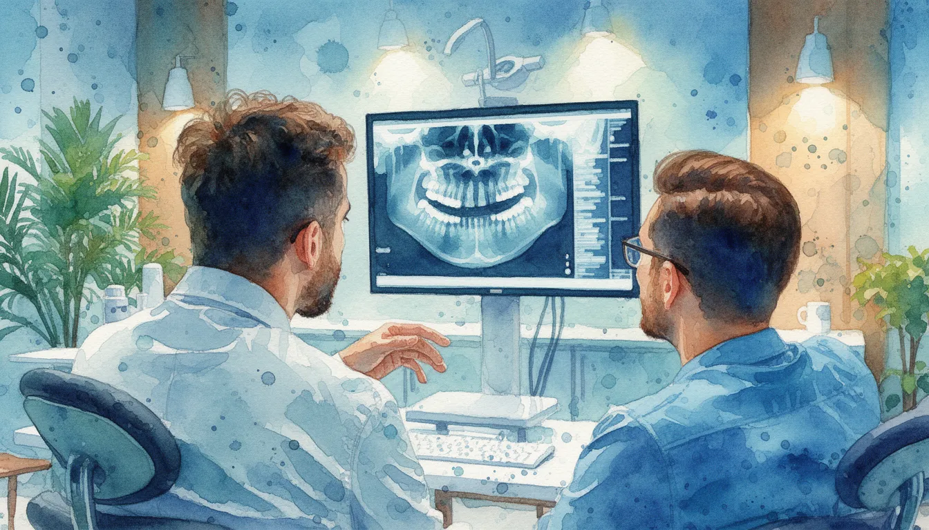
Unlike intraoral X-rays, where the film or sensor is placed inside your mouth, a panoramic X-ray is taken with the machine outside your mouth. The X-ray source and detector rotate around your head in a semicircle while you stand still. The result is a single image that your dentist can use to evaluate your overall dental and skeletal anatomy at a glance.
Panoramic X-rays have been used in dentistry since the 1960s and remain one of the most frequently ordered dental images. They serve as an excellent screening and diagnostic tool, though they do have limitations in resolution compared to smaller, more focused X-rays.
What a Panoramic X-Ray Shows
A single panoramic image reveals a wide range of dental and anatomical information. Your provider uses it to see the big picture before ordering more targeted imaging if needed.
Structures Visible on a Panoramic X-Ray
- All erupted and unerupted teeth, including wisdom teeth and their position relative to nerves
- The mandible (lower jaw) and maxilla (upper jaw), including overall bone height and density
- The temporomandibular joints (TMJs) on both sides
- The maxillary sinuses above the upper teeth
- The inferior alveolar nerve canal, which runs through the lower jaw
- Signs of jaw fractures, cysts, tumors, or other bone pathology
- The nasal cavity and surrounding bony structures
- Existing dental work, including fillings, crowns, implants, and root canal treatments
Common Clinical Uses
Dentists order panoramic X-rays for a variety of diagnostic and planning purposes. Wisdom tooth evaluation is one of the most common reasons. The panoramic view shows whether wisdom teeth are impacted, angled, or positioned near the inferior alveolar nerve. Orthodontic treatment planning uses panoramic images to count teeth, check for missing or extra teeth, and assess root length and jaw symmetry.
Other uses include screening for jaw cysts and tumors, evaluating bone loss from periodontal disease, planning dental implant sites (though CBCT is often preferred for precise implant planning), assessing facial fractures after trauma, and monitoring dental development in children and adolescents.
What to Expect During a Panoramic X-Ray
A panoramic X-ray is a quick, painless procedure. Understanding the process can help ease any concerns, especially for first-time patients or children.
Preparation
No special preparation is needed. You will be asked to remove any metal items from the head and neck area, including earrings, necklaces, eyeglasses, hearing aids, and removable dental appliances such as retainers or partial dentures. Metal objects can create artifacts on the image that obscure important details.
You may be given a lead apron or thyroid collar to wear during the scan, depending on the office's protocol. A thyroid collar protects the thyroid gland from scatter radiation, though the dose from a panoramic X-ray is already very low.
During the X-Ray
You will stand in front of the panoramic machine and place your chin on a small rest. You will bite gently on a small plastic tab that helps position your teeth correctly. The technician will adjust the machine to align with your anatomy.
Once positioned, you must remain still. The machine's arm will rotate around your head in a slow semicircle. The entire exposure takes about 15 to 20 seconds. You will not feel anything. The machine makes a quiet buzzing or humming sound as it moves. Some offices ask you to press your tongue flat against the roof of your mouth to reduce a shadow that can appear over the upper teeth.
After the X-Ray
The image is available almost instantly on a digital screen. Your dentist will review it with you, pointing out relevant findings. There is no recovery time. You can eat, drink, and resume all normal activities immediately.
Radiation Safety and Limitations
Understanding the radiation dose and the limitations of panoramic imaging helps you be an informed patient.
Radiation Exposure
A panoramic X-ray delivers a very low radiation dose, typically between 10 and 25 microsieverts (uSv). For perspective, the average person receives about 8 uSv per day from natural background sources like cosmic rays and radon gas. Flying cross-country exposes you to about 30-40 uSv. A single panoramic X-ray is roughly equivalent to one to two days of normal environmental radiation.
Modern digital panoramic machines use lower radiation doses than older film-based systems. The ALARA principle (As Low As Reasonably Achievable) guides all dental imaging decisions, meaning your provider should only order X-rays when there is a clear clinical reason.
Limitations of Panoramic X-Rays
Panoramic X-rays trade fine detail for broad coverage. They are excellent for seeing the big picture but have lower resolution than intraoral X-rays. Small cavities between teeth, early root fractures, and subtle periodontal bone changes may not be visible on a panoramic image.
Panoramic X-rays also produce some geometric distortion. Objects closer to the X-ray source appear magnified, and structures can overlap in ways that make interpretation challenging. Measurements taken from a panoramic X-ray are approximate, not exact. This is one reason CBCT scans are preferred for precise measurements, such as in implant planning.
When a CBCT Scan Is a Better Choice
A panoramic X-ray is a two-dimensional image. A CBCT scan produces a three-dimensional volume. If your provider needs to measure exact bone dimensions, see the precise relationship between a tooth and a nerve, or evaluate a lesion in 3D, a CBCT is the better tool. Panoramic X-rays are often the first step, and CBCT is ordered when more detail is needed.
Cost Factors for Panoramic X-Rays
Panoramic X-rays are among the most affordable dental imaging options. Costs vary by location and provider.
Typical Cost Range
A panoramic dental X-ray typically costs between $25 and $150 without insurance. Many dental offices include it as part of a new patient exam package. The price depends on your geographic area, the type of practice, and whether the image is digital (which is now standard in most offices).
Insurance Coverage
Most dental insurance plans cover panoramic X-rays. Many plans allow one panoramic X-ray every three to five years as part of routine care. Some plans cover it more frequently when a specific clinical need is documented, such as orthodontic treatment planning or evaluation of an injury. Check your plan's frequency limitations to avoid unexpected out-of-pocket costs.
When to See an Oral Radiology Specialist
Most panoramic X-rays are taken and interpreted by general dentists or dental specialists as part of routine clinical care. An oral and maxillofacial radiologist adds value in situations where interpretation is complex or findings are unexpected.
When a Radiologist Review Helps
- An unusual or suspicious finding appears on the panoramic image, such as a cyst, tumor, or calcification
- Your provider wants a second opinion before recommending surgery or additional imaging
- The image shows something outside the dental area, such as a sinus abnormality or a finding in the cervical spine
- You have a complex medical history that makes image interpretation more challenging
- A formal written radiology report is needed for referral, insurance, or legal purposes
Finding an Oral Radiology Specialist
Oral and maxillofacial radiologists are dentists who have completed a specialty residency focused on imaging interpretation, radiation physics, and diagnostic imaging techniques. They represent one of the 12 recognized dental specialties.
The American Academy of Oral and Maxillofacial Radiology (AAOMR) maintains a directory of specialists. Many oral radiologists offer teleradiology services, meaning your panoramic image can be uploaded and interpreted remotely. Ask your dentist whether they use a radiologist for image interpretation, especially if an unexpected finding appears on your panoramic X-ray.
Search Oral Radiologists in Your Area