What This Guide Covers and Who It Is For
This guide explains how successful gum grafts are, based on graft type, technique, and patient factors. It is written for anyone considering gum graft surgery or trying to understand their options.
Gum recession is the process where gum tissue pulls back from the tooth, exposing the root surface underneath. When enough root is exposed, it can cause sensitivity, increase the risk of decay, and affect the appearance of your smile. Gum grafting is a surgical procedure that covers exposed roots and rebuilds lost tissue. [1]
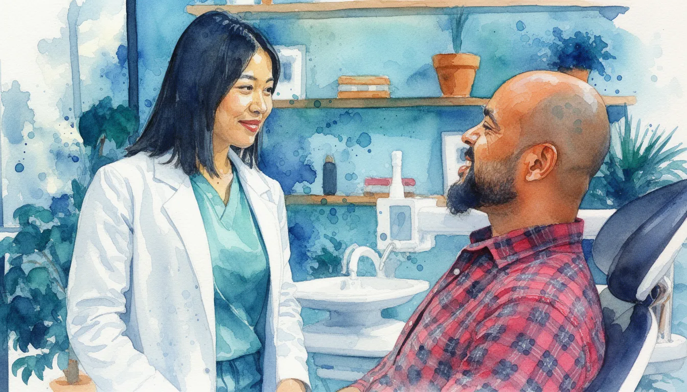
A periodontist is a dentist who has completed additional years of training focused on the gums, bone, and supporting structures of the teeth. These specialists perform gum grafts regularly and select the graft type based on the severity of recession, the thickness of your existing tissue, and your overall oral health. [1]
The sections below walk through what research shows about success rates for each graft type, what factors raise or lower those rates, what recovery looks like, and how costs typically break down.
Gum Graft Success Rates by Type
Success rates depend on the type of graft used, the severity of recession, and patient health factors like smoking status. It is important to understand two different ways researchers measure graft success: the average percentage of the exposed root that gets covered (mean root coverage) and the percentage of cases where every bit of exposed root is covered (complete root coverage). These are different numbers, and knowing the difference helps you set realistic expectations.
Connective Tissue Graft (CTG)
The connective tissue graft is the most studied and most commonly recommended option for root coverage. In this procedure, a small piece of tissue is taken from beneath the surface layer of the palate (the roof of the mouth) and placed over the exposed root.
Published research consistently shows that connective tissue grafts achieve high levels of root coverage on Miller Class I and Class II recession defects, where the bone between the teeth is still intact. A 2008 systematic review by Cairo et al., which analyzed multiple controlled clinical trials, found that CTGs produced a mean root coverage of approximately 90%, meaning that on average about 90% of the exposed root surface was covered. The same review found that complete root coverage, where 100% of the exposed root is covered, was achieved in approximately 60% of treated sites for Miller Class I and II defects. [4] Even when complete coverage is not achieved, most patients still see a significant reduction in exposed root surface.
Periodontists often call the CTG the "gold standard" because it consistently produces thick, stable tissue that blends well in color with the surrounding gums over time. The tissue taken from beneath the palate surface tends to heal with a good blood supply, which supports long-term stability of the graft. A separate systematic review by Tavelli et al. has provided important anatomical guidance on safe tissue harvesting from the palate, helping periodontists minimize complications at the donor site. [3]
Free Gingival Graft (FGG)
A free gingival graft takes a thin piece of tissue directly from the surface of the palate and places it at the gum line. Unlike the CTG, this graft includes the outer epithelial layer (the visible surface layer of tissue).
Free gingival grafts are not typically used when the primary goal is root coverage. Their root coverage rates are notably lower than those of connective tissue grafts. However, they excel at a different task: building a wider band of thick, attached gingiva (firm gum tissue that does not move). This is especially useful around teeth or implants where the existing gum tissue is very thin or fragile.
Because the tissue comes from the surface of the palate, the color match with surrounding gums is sometimes less natural compared to CTG results. The graft site on the palate also tends to heal more slowly, since the tissue is removed directly from the surface rather than from underneath it.
Allografts, Xenografts, and Collagen Matrices
Allografts use donated human tissue from a tissue bank. Xenografts use processed tissue from animal sources. Collagen matrices are manufactured products designed to act as a scaffold for your own tissue to grow into. All three eliminate the need for a second surgical site in your mouth.
These alternatives are attractive to patients because they reduce surgical time and avoid palate discomfort. Research suggests they can produce good root coverage results, though many studies show slightly lower complete root coverage rates compared to connective tissue grafts taken from the patient's own palate. A 2015 systematic review and meta-analysis by Chambrone et al., which pooled data from multiple randomized controlled trials, confirmed that autogenous connective tissue grafts (tissue from your own body) provided statistically superior root coverage compared to substitute materials, though substitute materials still achieved clinically meaningful results. [5]
Your periodontist may recommend one of these options when there is not enough palate tissue available for a traditional graft, when multiple teeth need treatment at the same time, or when you prefer to avoid a palate donor site. The choice involves weighing the potential for slightly different outcomes against the benefit of a less invasive procedure.
How Miller Classification Affects Success
The Miller classification system grades recession defects from Class I to Class IV based on how much bone and tissue has been lost between the teeth. It is the strongest predictor of how much root coverage any graft can achieve.
Miller Class I and Class II defects still have intact bone between the teeth (called interdental bone). These cases have the highest success rates. As noted above, complete root coverage is achievable in roughly 60% of these cases when a connective tissue graft is used, with average root coverage around 85% to 95%. [4] Miller Class III defects involve some loss of interdental bone or slight tooth malpositioning, so only partial root coverage is typically realistic. Miller Class IV defects involve severe bone loss, and complete root coverage is generally not predictable with any technique.
Your periodontist will measure your recession and assess the bone level before surgery. This classification directly shapes the treatment plan and helps set realistic expectations for how much coverage you can expect.
Factors That Affect Your Graft Outcome
Several controllable and uncontrollable factors influence how well a gum graft heals and how much root coverage it ultimately achieves.
Smoking and Tobacco Use
Smoking is the single biggest controllable risk factor for gum graft failure. Tobacco restricts blood flow to the gums, which directly interferes with the healing process that a graft depends on.
Some clinical studies have reported that smoking reduces root coverage outcomes by 25% or more. A systematic review by Chambrone et al. found that smoking was a significant negative predictor of root coverage success across multiple graft types. [5] Many periodontists ask patients to stop smoking for at least two weeks before and after surgery to give the graft the best chance of survival. If quitting entirely is not possible, even reducing the amount you smoke during the healing window can make a meaningful difference.
Tissue Thickness and Gum Biotype
People naturally have different gum thicknesses. A thicker gum biotype (sometimes called a thick-flat biotype) tends to heal more predictably after grafting. Thin, delicate tissue is more fragile during surgery and during the early healing phase.
Your periodontist will evaluate your tissue thickness during the initial exam. In cases where the tissue is very thin, the surgeon may choose a technique specifically designed to add volume, such as a free gingival graft for thickening before or instead of a root coverage procedure.
Oral Hygiene Before and After Surgery
Plaque and bacteria around the surgical site can compromise graft healing. Your periodontist will typically recommend a professional cleaning before the procedure to reduce bacterial load. [1]
After surgery, you will receive specific instructions about how to keep the area clean without disturbing the graft. This usually involves avoiding brushing directly on the graft site for one to two weeks and using a prescribed antimicrobial mouth rinse instead. Following these instructions closely has a direct impact on the outcome.
Age and Timing Considerations
Gum grafting can be performed on adults of any age. There is no upper age limit, as long as the patient is healthy enough for minor oral surgery. Younger patients sometimes heal faster, but older adults with good health routinely achieve successful outcomes.
Timing matters. Recession that is caught early, before significant bone loss occurs, is easier to treat and more likely to achieve complete root coverage. If you notice your gums pulling away from a tooth, or if a tooth looks longer than it used to, scheduling a periodontal evaluation sooner rather than later gives you more treatment options.
Understanding How Success Is Measured
When reading about gum graft success rates, it helps to know that researchers use two main measurements. Mean root coverage refers to the average percentage of exposed root that gets covered across all treated sites. Complete root coverage (CRC) refers to the percentage of cases where every bit of exposed root is covered, a perfect result. Mean root coverage numbers are typically higher than CRC rates because even partial coverage counts toward the average. [4]
For example, a study might report a mean root coverage of 90% and a complete root coverage rate of 60%. This means the average patient had about 90% of their exposed root covered, but only about 60% of patients had every last bit of root covered. Both numbers reflect good outcomes, but understanding the difference helps you have a more informed conversation with your periodontist about what to expect.
What to Expect Before, During, and After Surgery
Gum graft surgery is typically performed in one visit under local anesthesia, and most patients return to normal activities within a few days.
Before the Procedure
Your periodontist will take detailed measurements of the recession, examine your palate as a potential donor site, and review your medical history. X-rays or a cone beam scan may be taken to evaluate bone levels around the affected teeth.
You will receive instructions about medications to avoid before surgery (such as blood thinners, if medically safe to pause). If sedation is planned beyond local anesthesia, you will need someone to drive you home.
During the Procedure
The procedure typically takes 60 to 90 minutes, depending on how many teeth are being treated. Your periodontist will numb the area with local anesthesia. You should feel pressure but not pain during the surgery.
For a connective tissue graft, the surgeon makes a small flap in the palate, removes a piece of connective tissue from underneath, and then closes the palate flap with sutures. The donor tissue is placed over the exposed root and secured with tiny sutures. Research on palatal anatomy has mapped safe harvesting zones to help periodontists avoid the greater palatine artery during this step, reducing the risk of excessive bleeding. [3] For a free gingival graft, the tissue is taken directly from the palate surface, leaving an open wound that heals on its own over several weeks.
Recovery and Healing Timeline
Most patients experience mild to moderate discomfort for the first two to three days. Pain is usually manageable with over-the-counter pain relievers or a short prescription. Swelling and some bruising are normal.
Initial healing of the graft site typically occurs within two to three weeks. During this time, you will eat soft foods and avoid chewing near the graft. The palate donor site, if applicable, usually takes two to four weeks to feel comfortable again.
Final tissue maturation takes longer. The graft tissue gradually blends in color and texture with the surrounding gums over three to six months. During follow-up visits, your periodontist will monitor the graft attachment, tissue thickness, and the amount of root coverage achieved.
Gum Graft Cost and Insurance Considerations
Gum graft costs vary widely depending on the graft type, number of teeth treated, and geographic location.
As a general range, a gum graft for a single tooth may cost between $600 and $3,000. When multiple teeth are treated in one session, the per-tooth cost may decrease, but the total cost rises. Procedures that use allograft or collagen matrix materials may carry additional material fees. Costs vary by location, provider, and case complexity.
Many dental insurance plans classify gum grafting as a medically necessary periodontal procedure rather than a cosmetic one, especially when recession is causing sensitivity, decay risk, or progressive tissue loss. Coverage levels vary by plan. Some plans cover 50% to 80% of the procedure cost after deductibles, while others may apply annual maximums that limit the benefit. [2]
Before scheduling, ask your periodontist's office for a pre-treatment estimate that can be submitted to your insurance company. This gives you a clearer picture of your out-of-pocket responsibility. Many offices also offer payment plans or financing options to help spread the cost over time.
When to See a Periodontist vs. a General Dentist
A periodontist should evaluate your case when recession is progressing, causing symptoms, or when grafting is being considered as a treatment option.
General dentists are skilled at identifying gum recession during routine exams. Many will refer you to a periodontist when the recession reaches a point where surgical treatment may be beneficial. You can also seek a periodontal evaluation on your own if you have noticed changes in your gum line. [1]
Consider scheduling a periodontal consultation if you notice one or more of the following: a tooth that appears longer than neighboring teeth, visible root surface at the gum line, increased sensitivity to hot or cold along the gum line, a notch you can feel with your fingernail where the gum meets the tooth, or if your general dentist has mentioned thinning gums during a checkup.
A periodontist has specialized training in gum tissue surgery and will evaluate the Miller classification of your recession, your tissue thickness, your bone levels, and your overall risk factors. This evaluation is the foundation for recommending a specific graft type and setting realistic expectations for root coverage. You can learn more about what this specialist does on the periodontics page.
Find a Periodontist Near You
If you are experiencing gum recession or have been told you may need a gum graft, a periodontist can evaluate your specific situation and recommend the graft type most likely to produce a good result. Use the search tool on the periodontics page to find a qualified periodontist in your area and schedule a consultation.
Search Periodontists in Your Area